Quantitative Phase Microscopy (QPM) is a recently developed computational approach that provides quantitative phase measurements of images captured using a bright-field microscope. Phase measurement is particularly useful in the evaluation of translucent objects, such as unstained viable cell specimens. QPM works via an algorithm which is applied to a standard bright-field and equidistant positive and negative de-focus images. From these images a phase map is generated which contains information about cell thickness and refractive index and can allow quantitation of cellular structure. QPM was used in conjunction with laser scanning confocal microscopy (LSCM) to measure volume and area in cultured human airway smooth muscle (HASM) cells.
HASM cells were obtained by collagenase and elastase digestion of smooth muscle from lung transplant resection patients. The resulting cell suspension was washed in phosphate buffered saline and seeded onto glass coverslips which were placed in the base of a plastic culture dish at 37°C in Dulbecco's Modified Eagles Media. The cells were imaged using both LSCM (optical slicing) and QPM techniques. For LSCM visualisation, cells were fluorescently labelled (fluo-3/AM, Molecular Probes, Eugene, OR, USA) and a series of images in the vertical (z) axis were recorded at intervals of 1μm using a Leica TCS 4D (x63, PL APO 1.40 NA oil immersion objective). From these images a calculation of cell depth was performed. Phase images of the same field of cells were then obtained using an inverted Zeiss Axiovert 100M microscope (×10, LD-Achroplan 0.3 NA objective) and a Coolsnap fx CCD camera (Photometrics, USA). Phase calculations were performed using QPm software (v2.0 IATIA Ltd, Australia).
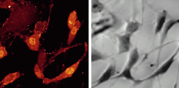
On the basis of the cell depth determined by LSCM (left panel of Figure) and the phase measurements calculated by QPM (right panel of Figure) a mean refractive index (RI) for HASM was determined to be 1.4275 ± 0.009. This RI was then used computationally by the QPM algorithm to determine cellular volume. The relationship between cell confluency and volume in HASM cultures passaged for variable periods was evaluated. The confluency of cells (% field area), calculated over a 92 hour growth period from phase maps and cell volume (μm3) were highly correlated (i.e. r2 values of 0.986 and 0.996 obtained for two different cell culture lines). Thus, in these HASM cells under the conditions specified, there is a well defined relationship between extent of confluence and total cell volume in culture. QPM provides a convenient procedure for estimating cell volume, where prior determination of RI is required. We have demonstrated that this may be achieved by parallel specimen imaging using both LSCM and QPM.