The role of Ca2+-release from the sarcoplasmic reticulum in influencing the pacemaker rate appears to be a common mechanism in sinuatrial node (SA) (Hata et al., 1996) and atrioventricular node (AV) (Hancox et al., 1994). An increased level of intracellular Ca2+ or Ca2+ overload has been suggested to be a major cause of arrhythmia (Blaustein & Lederer, 1999). Calbindin D-28K and parvalbumin calcium-binding proteins have been found in neurons, and function as intracellular Ca2+ buffering transport protein to maintain a constant intracellular Ca2+ level (Chard et al., 1993). However, there have been no previous reports of calcium-binding protein in the SA and AV nodes. This study aimed to investigate whether the SA and AV nodes contain calbindin D-28K and parvalbumin.
Adult Wistar rats age 4 weeks were anaesthetised by ether inhalation. The hearts were removed and fixed in 10% formaldehyde. Following dehydration and embedding in paraffin, 5μm serial sections were cut in both the horizontal and coronal planes and then stained with Masson Trichrome to localise the SA and AV nodes. The sections containing SA or AV node were processed for immunohistochemical labeling with antibodies to calbindin D-28K and parvalbumin.
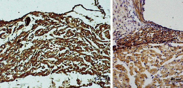
(A) Cross section of the heart at the junction of superior vena cava and right atrium, stained with calbindin D-28K antibody at a dilution of 1:400, showing immunoreactivity (brown color) in the cytoplasm of nodal fibers of SA node. (B) Coronal section of the heart,stained with parvalbumin antibody at a dilution of 1:2000, showing immunoreactivity (brown color) in the cytoplasm of nodal fibers of AV node.
The calbindin D-28K and the parvalbumin are found in both the SA and AV nodes, suggesting these two binding proteins may be involved in the generation and conduction of electrical impulses by maintaining a constant level of the intracellular calcium ions. It is hoped that the study of these calcium binding proteins will open new avenues for therapeutic interventions, especially for the treatment of Ca2+ overload and arrhythmia.
Blaustein, M.P. & Lederer, W.J. (1999) Physiological Review, 79, 763-854.
Chard, P.S., Bleakman, D., Christakos, S., Fullmer, C.S. & Miller, R.J. (1993) Journal of Physiology, 472, 341-357.
Hancox, H., Lederer, W.J. & Cannell, M.B. (1993) Proceedings of the Royal Society B, 255, 99-105.
Hata, T., Noda, T., Nishimura, M. & Watanabe, Y. (1996) Heart and Vessels, 11, 234-241.