Background: Brain-derived neurotrophic factor (BDNF) is a potent molecule regulating dendritic trees and synaptic plasticity, however, whether the BDNF precursor (proBDNF) plays any role in the neurite growth is unknown. This study investigated whether proBDNF has a physiological role in the neurite growth of neonatal cortex neuron in vitro. We focus on the following the aspects, the dose effect of proBDNF on neurite outgrowth, ELISA assay to test proBDNF and BDNF after stimulated neurons by 45 mM KCl for 30 min, distribution of RohA, cdc42, F-actin via immunohistochemistry after gradient or uniform of proBDNF added, and examined the down streaming signals of RhoA and cdc42.
Methods: The cortex from neonatal C57BL/6 (n=12) mice were rapidly removed and cultured. Live images were collected every 6 s for 30 min by using Nikon BioStation and analyzed via the advanced software. Primary neurons were incubated in proBDNF at various concentrations for 24 h and the lengths were measured. 8 × 10⁶ cells were dissociated for 24 h, treated with 50 ng/ml proBDNF for 0, 5, 10, 20 min, the neuron cell lysates of 100 μg protein in 0.5 ml RIPA buffer were incubated in 10 μg Rho binding domain (RBD) agarose gel for RhoA and cdc42 activity assays. The pull-down and total proteins were evaluated by western blotting. Data are mean ± SEM from 3 independent experiments.
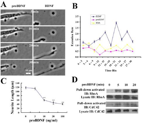
Results: Live imaging showed the clear collapse of growth cones in response to proBDNF (50 ng/ml) and the elongation to BDNF (50 ng/ml) at the indicated point (Figure A). proBDNF caused a 34% decrease in the extension rate after 6 min and maintained throughout a 30 min treatment. Meanwhile, neurons treated with BDNF spent 63% of the time extending (Figure B). proBDNF (10, 30, 100 ng /ml) resulted in a 50% decrease in length (58.6 μm vs 118.1 μm) compared with 0 ng /ml proBDNF group (Figure C) but proBDNF 3 ng /ml had no significant effects on the length. 5 min after proBDNF treatment, RhoA activity was increased by 5 fold 20 min after proBDNF incubation (Figure D). Interestingly, no difference in activated cdc42 level was seen during the proBDNF incubation.
Conclusion: Live imaging demonstrated that proBDNF repulsed growth cones and induced the neurite collapse of cortical neurons in neonatal mice. Statistical analysis showed the dose-dependent effect of proBDNF on neurite collapse. proBDNF rapidly activated RhoA without affecting the cdc42 level. Thus, proBDNF inhibits neurite growth via activating RhoA in neonatal cortical neurons of mice.