In specialized cells, fusion of secretory vesicles with the plasma membrane enables release of biologically active molecules; this process is tightly regulated by intracellular Ca2+. However, the identity of critical proteins involved in Ca2+ sensing and triggering of native membrane fusion remain speculative. Unlike other types of secretory vesicles, isolated cortical vesicles (CV) from unfertilized urchin eggs remain fully primed and fusion-ready providing a stage-specific preparation to quantitatively assess the native, Ca2+-triggered fusion mechanism (Zimmerberg et al., 2000). Thiol-reactivity offers an unbiased approach in studying proteins involved in Ca2+-triggered membrane fusion. Furthermore, alkylating reagents are highly selective for, and bind irreversibly to, cysteine residues, providing important information about function while also tagging the proteins involved.
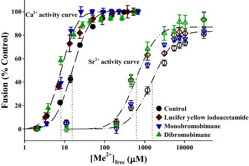
Previous work indicates that multiple thiol sites differentially regulate the ability of CV to fuse as well as the efficiency of fusion (i.e. Ca2+ sensitivity and kinetics) (Furber, Brandman & Coorssen, 2009; Furber, Dean & Coorssen, 2010). Iodoacetamides and bimanes have the unique ability to access a novel thiol site that potentiates the efficiency of fusion, presumably by altering the Me2+ sensing mechanism (Figure).
Labeled proteins thus far identified include a variety of metabolic enzymes, cystoskeletal components (actin and tubulin), and several isoforms of Rab GTPases. Both the cystoskeleton and Rab proteins have defined roles in vesicle mobility and trafficking, but these data raise the possibility they may also act in later stages of exocytosis to regulate Ca2+ sensitivity and kinetics of secretion. Pharmacological experiments using cytoskeletal (de)stabilizing reagents in the stage-specific CV preparation rule out a direct role for actin (Hibbert, Butt & Coorssen, 2006) and tubulin (Furber et al., unpublished) in the Ca2+-triggering steps of membrane fusion. Nonetheless, there is evidence indicating a role for cytoskeletal components in fusion pore expansion and thus the kinetics of the release process (Berberian et al., 2009; Doreian, Fulop & Smith, 2008; Larina et al., 2007; Miklavc et al., 2009). We are currently focusing on Rab proteins as prime candidates involved in the regulation of fusion efficiency, perhaps reminiscent of the synergistic effects between Ca2+ (CE) and GTP (GE) on secretion in other cell types (Coorssen, Davidson & Haslam, 1990; Howell, Cockcroft & Gomperts, 1987).
Berberian K, Torres AJ, Fang Q, Kisler K & Lindau M (2009) Journal of Neuroscience, 29, 863-870.
Coorssen JR, Davidson MM & Haslam RJ (1990) Cell Regulation, 1, 1027-1041.
Doreian BW, Fulop TG & Smith CB (2008) Journal of Neuroscience, 28, 4470-4478.
Furber KL, Brandman DM & Coorssen JR (2009) Journal of Chemical Biology, 2, 27-37.
Furber KL, Dean KT & Coorssen JR (2010) Clinical and Experimental Pharmacology and Physiology, 37, 208-217.
Hibbert JE, Butt RH & Coorssen JR (2006) International Journal of Biochemistry and Cell Biology, 38, 461-471.
Howell TW, Cockcroft S & Gomperts BD (1987) Journal of Cell Biology, 105, 191-197.
Larina O, Bhat P, Pickett JA, Launikonis BS, Shah A, Kruger WA, Edwardson JM & Thorn P (2007) Molecular Biology of the Cell, 18, 3502-3511.
Miklavc P, Wittekindt OH, Felder E & Dietl P (2009) Annals of the New York Academy of Science, 1152, 43-52.
Zimmerberg J, Blank PS, Kolosova I, Cho MS, Tahara M, & Coorssen JR (2000) Biochimie, 82, 303-314.