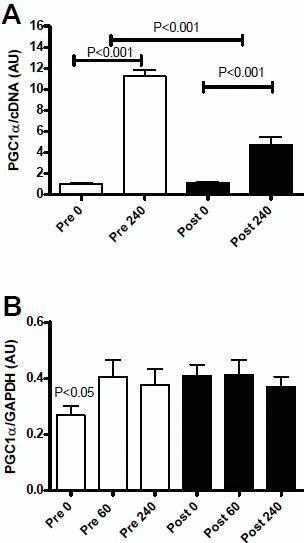
Mitochondrial biogenesis and function are important for energy production in cells and tissues. Aberrant mitochondrial function, specifically reduced volume not function, has been implicated as a cause or at least a contributor to lifestyle diseases including insulin resistance, obesity and diabetes. It is also well established that mitochondrial function and biogenesis is promoted by physical activity and exercise. In this study we investigated whether mitochondrial biogenesis was maintained in response to acute exercise after 10d of intensive cycle training despite the reduction of AMPK activity. Nine untrained, healthy participants (mean ± SEM; 23 ± 5 years of age, BMI: 24.9 ± 1 kg.m−2 VO2peak 44.1 ± 7.2 ml.kg−1.min−1) provided written informed consent. These participants performed a 60 min bout of cycling exercise at 164 ± 9 W (∼70% pre-training VO2peak), muscle biopsies were taken from the vastus lateralis muscle under local anesthesia at rest, immediately and 3h after exercise. Within 7 days the participants then underwent 10d of intensified cycle training including 4 days of high-intensity interval training. Three days after the final training session participants repeated the pre-training exercise trial with biopsies at the same absolute work load (∼164 W). Protein and mRNA were extracted from muscle for analysis by immunoblotting or RT QPCR respectively. AMPK Thr172 phosphorylation increased by 15 fold and 4 fold during exercise before and after training respectively (p<0.05). PGC1-α gene expression was increased by 11 and 4 fold (p<0.001; Figure A) 3 h after the exercise bout before and after training.