Enteroendocrine cells located in the mucosal layer throughout the gastrointestinal tract constitute the largest endocrine organ in the human body. Amongst this the largest single population of cells are the enterochromaffin (EC) cells which produce ∼95% of the body's serotonin (5-hydroxytryptamine or 5-HT) (Erspamer, 1954). This circulating 5-HT is vital for a multitude of bodily functions including enteric motility (Keating & Spencer, 2010), bone mass (Yadav et al., 2008), liver regeneration (Lesurtel et al., 2006) and haemostasis (Walther et al., 2003). Dysfunctional 5-HT release from EC cells has been implicated in human health disorders including irritable bowel syndrome, Crohn's disease and osteoporosis (Gershon & Tack, 2007). Understanding how 5-HT is released from EC cells and the mechanisms that control this release is therefore important in terms of both health and disease. In spite of their importance, no study has yet examined 5-HT release from single primary EC cells.
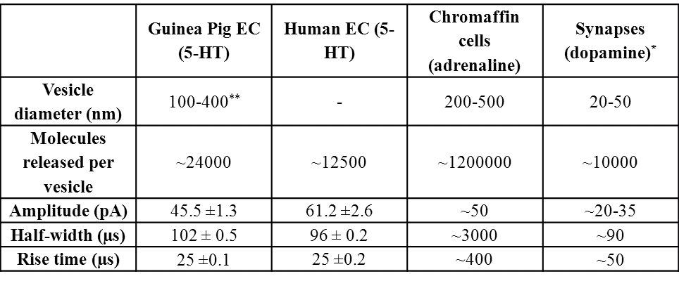
This study presents for the first time a rapid method of isolating and purifying (∼99% 5-HT-positive) guinea pig and human EC cells in primary culture. We used carbon fibre amperometry to measure 5-HT release from single EC cells and find that they release ∼60-100 times less 5-HT per vesicle fusion event compared to other endocrine cells and that the kinetics of 5-HT release resembles that occurring in synapses (the Table). Thus 5-HT may be released from EC cells via an extremely fast “kiss and run” type of exocytosis not previously observed in endocrine cells. We also identify a range of endogenous factors including acetylcholine, glucose and 5-HT itself which stimulate calcium entry and 5-HT release from EC cells. These findings are the first study demonstrating the basic properties underlying stimulus-secretion coupling in EC cells. This includes a mode of exocytosis unique amongst all reported endocrine cells and suggests a novel paradigm in neurotransmitter release studies.
Erspamer, V. (1954) Pharmacological Reviews 6, 425-487.
Gershon, M.D. and Tack, J. (2007) Gastroenterology 132, 397-414.
Keating, D.J. and Spencer, N.J. (2010) Gastroenterology 138, 659-670.
Lesurtel, M., Graf, R., Aleil, B., Walther, D.J., Tian, Y., Jochum, W., Gachet, C., Bader, M. and Clavien, P.A. (2006) Science 312, 104-107.
Walther, D.J., Peter, J.U., Winter, S. Höltje M, Paulmann N, Grohmann M, Vowinckel J, Alamo-Bethencourt V, Wilhelm CS, Ahnert-Hilger G, Bader M. (2003) Cell 115, 851-862.
Yadav, V.K., Ryu, J.H., Suda, N. Tanaka KF, Gingrich JA, Schütz G, Glorieux FH, Chiang CY, Zajac JD, Insogna KL, Mann JJ, Hen R, Ducy P, Karsenty G. (2008) Cell 135, 825-837.