Control of blood flow and pressure involves maintaining a normal balance of vasoconstrictor and vasodilator activity, as artery tone, which is altered in vascular disease. Endothelial cells lining the vessel lumen contribute to tone via their production of constrictor and dilator agents, with key aspects of artery function being dependent on microdomain signaling and the associated modulation of intracellular calcium, with transient receptor potential (TRP) channels playing an integral role (Sandow et al. 2012; Senadheera et al. 2012). This study determines the distribution and role of TRP canonical type-3 (C3), in the control of endothelium-derived hyperpolarization (EDH)-mediated vasodilator tone in rat mesenteric artery, as a model resistance vessel, where intermediate conductance calcium activated potassium channels (IKCa) and endoplasmic reticulum (ER) inositol 1,4,5-trisphosphate receptor (IP3R) microdomains are critical.
TRPC3 antibody specificity was verified using rat tissue, HEK-293 cells stably transfected with mouse TRPC3 cDNA, and TRPC3 knock-out mouse tissue using Western blotting, confocal and ultrastructural immunohistochemistry. TRPC3-Pyr3 blocker specificity was verified using patch clamp of endothelial and TRPC3-transfected HEK cells, and TRPC3 knock-out and wild-type mouse aortic endothelial cell calcium imaging and mesenteric artery pressure myography. TRPC3 distribution, expression and role in EDH-mediated function were examined in rat mesenteric artery using immunohistochemistry and Western blotting, and pressure myography and endothelial cell membrane potential recordings. IKCa, IP3R and ER distribution were examined using confocal immunohistochemistry and electron microscopy. All procedures were approved according to respective Institutional ethics committees and National guidelines.
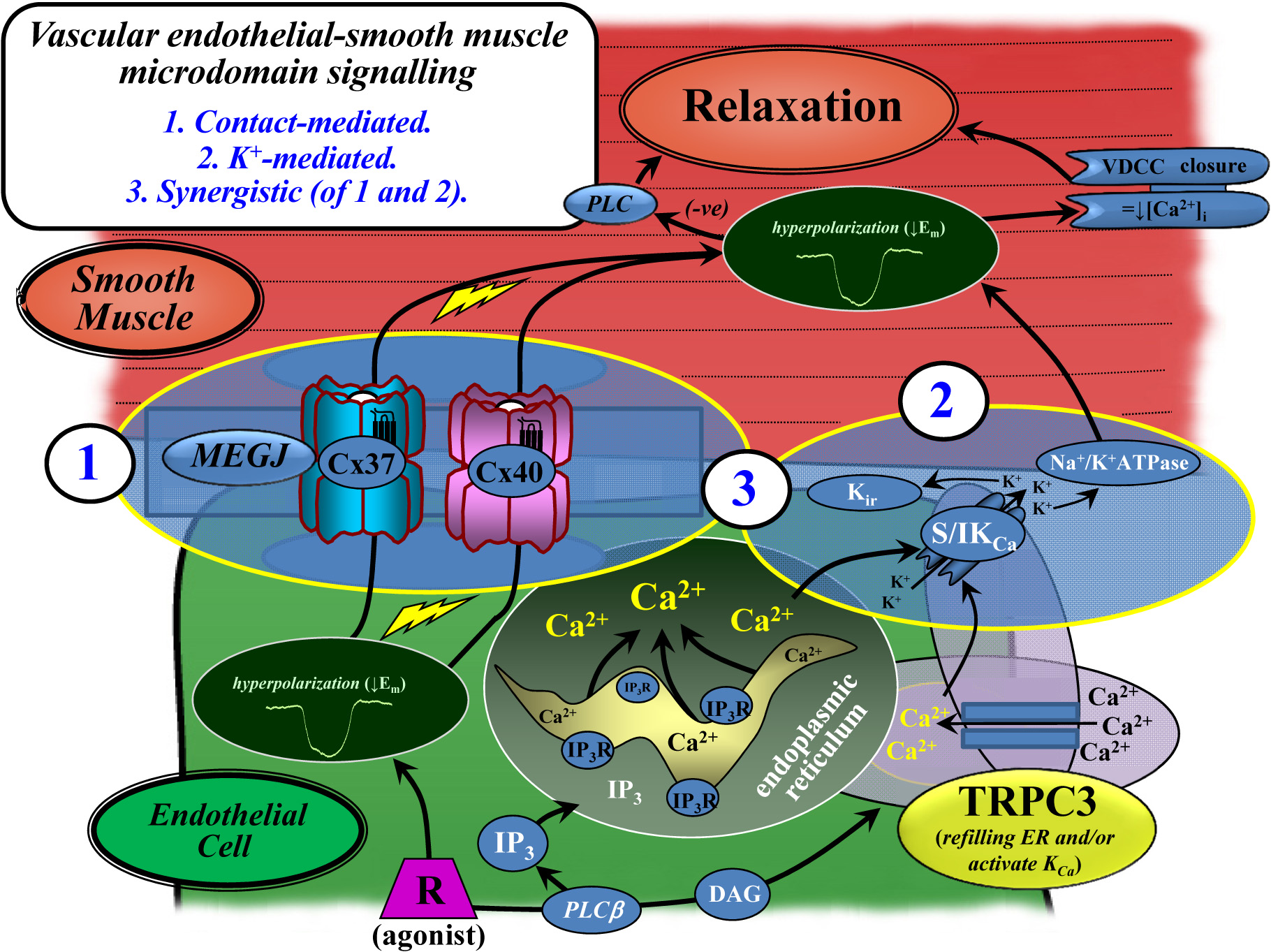
In rat mesenteric artery, TRPC3 is diffusely distributed in the endothelium, with ∼5-fold higher expression at myoendothelial microdomain contact sites; with immunoelectron microscopy confirming TRPC3 at these sites, where high level IKCa and ER-IP3R are also present. Western blotting confirmed primary endothelial TRPC3 expression. In rat mesenteric artery endothelium, Pyr3 inhibited EDH; and with individual SKCa (apamin) or IKCa (TRAM-34) block, Pyr3 abolished the residual respective IKCa and SKCa-dependent EDH-mediated vasodilation. TRPC3-IKCa and ER-IP3R-calcium stores are co-localized at discrete sites between the endothelium and smooth muscle.
TRPC3 facilitate endothelial SKCa and IKCa activation, as key components of the EDH-mediated rat mesenteric artery vasodilator mechanism (Figure). The integrated microdomain function of TRPC3 and SKCa-IKCa, and there spatial localization with one another and IP3R-ER calcium stores supports there role in the control of vascular tone tone and blood flow. Diversity in microdomain components are key targets for future investigation as factors underlying differential vessel function in health and disease.
Sandow SL, Senadheera S, Bertrand PP, Murphy TV, Tare M (2012) Microcirculation 19:403-415.
Senadheera S, Kim Y, Grayson TH, Toemoe S, Kochukov MY, Abramowitz J, Housley GD, Bertrand RL, Chadha PS, Bertrand PP, Murphy TV, Tare M, Birnbaumer L, Marrelli SP, Sandow SL (2012). Cardiovascular Research 95:439-447.