Pointillistic super-resolution imaging techniques can routinely achieve lateral spatial resolution that is an order of magnitude better than regular diffraction limited fluorescence imaging (200-300 nm). The underlying principle is that the position of an emitter can be determined very accurately if its emission pattern can be imaged unperturbed. This is achieved even with high labelling densities by temporal separation of the emitters via “switching” the molecules between being emissive and non-emissive or “dark” states. dSTORM or direct stochastic optical reconstruction microscopy achieves sub-diffraction imaging using conventional fluorescent probes, such as labelled antibodies, combining with a reducing medium so that the vast majority of fluorophores are shifted into a stable dark state. Stochastic recovery from this non-fluorescent state allows individual fluorphores to be imaged a few at a time and their spatial coordinates to be determined. By collating all of the fitted PSFs, a super-resolution image can be constructed.
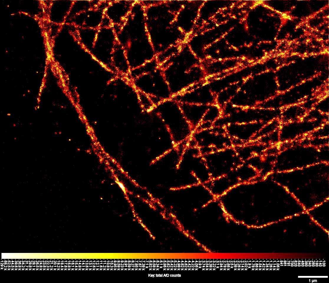
Here we report the recent construction and testing of a home built dSTORM super-resolution microscope built at Monash University from “off-the-shelf” components for less than A$100K. The instrument has so far been used generate super-resolution images of microtubules and actin filaments in fixed HeLa and COS-7 cells at better than 50 nm resolution. The Figure shows an example of microtubules labelled with Alexa-647 in a HeLa cell.