Contents


|
|
Programme
Contents
|
Insulin-stimulated glucose uptake is reduced in people with type 2 diabetes (T2D), especially in skeletal muscle, which accounts for >70% of insulin-stimulated whole-body glucose uptake. Importantly, however, skeletal muscle glucose uptake during exercise is normal in people with T2D and in rodent diabetic animal models. In addition, increases in insulin sensitivity with regular exercise training are normal in T2D. The signalling events responsible for exercise-induced enhancement of insulin-stimulated glucose uptake are unclear and represent an attractive target to improve glucose homeostasis in T2D. A large animal model with similar responses to exercise as seen in humans would facilitate further mechanistic studies of the effects of acute exercise and exercise training on insulin sensitivity, whilst allowing direct measurement of insulin action and repeated biopsies in longitudinal studies of exercise training effects.
Eight 6 yr old non-pregnant Border Leicester × Merino ewes were acclimatised to stand and walk slowly (2-3 minutes at 0.8 km/h, 0% slope) on a treadmill for three days prior to surgery and two days after surgery. PVC catheters were inserted into the left jugular vein and left carotid artery of each ewe under general anaesthesia and asepsis. Whole-body insulin sensitivity was measured by hyperinsulinaemic euglycaemic clamp (HEC,120 min, 2 mU insulin.min-1.kg-1) (Pre). Sheep were then trained to walk more briskly up a slope over ten days. On the 11th day they undertook an acute intensive treadmill exercise bout at 8° slope, consisting of a 3 min stepwise increase in speed from 0.8 to 4.0 km/h, then 15 min at 4.4 km/h, 3 min at 1.6 km/h and 15 min at 4.0 km/h. This exercise intensity equates to 50-60% of VO2 max, based on published data for pregnant sheep. A HEC was conducted ∼18 hours after the acute intensive exercise bout (Post). A M. semimembranosus biopsy was collected under local anaesthesia (1% Lignocaine) before and immediately following each HEC. All procedures were approved by the University of Adelaide Animal Experimentation and Ethics Committee.
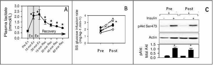
Plasma lactate concentration increased significantly (p < 0.05) during acute exercise and remained elevated during the recovery period (Figure panel A). Plasma glucose did not change during acute exercise or recovery. Acute exercise reduced fasting plasma glucose prior to the HEC (Pre: 3.7 ± 0.08 mmol.L-1, Post: 3.5 ± 0.1 mmol.L-1, p = 0.003) and the steady-state (SS) glucose infusion rate required to maintain euglycaemia during the HEC increased by 49% (Pre: 1.92 ± 0.12 mg.kg-1.min-1, Post: 2.85 ± 0.21 mg.kg-1.min-1, p = 0.003, Figure B) and insulin sensitivity increased by 65% (p = 0.018) after exercise. The HEC increased phosphorylation (activation) of Akt Ser473, indicative of proximal insulin signalling, by ∼5-fold, with no difference after exercise training (Figure panel C, p < 0.05), consistent with human responses.
Our findings of increased plasma lactate during exercise, and increased insulin sensitivity with unchanged insulin signalling the day after exercise are consistent with human studies and indicate that sheep are a suitable large mammal model to investigate underlying mechanisms for the beneficial effects of exercise on insulin sensitivity.