Background: The adult human heart has very little capacity to repair damage because adult cardiomyocytes lack the ability to proliferate; the immune system plays an important role in modulating this response. The immune system of fetuses possesses diminished pro-inflammatory function, and an immature immune system, which is believed to provide an immunological setting in which cardiomyocytes can regenerate.
Methods: We used sheep, which have similar cardiomyocyte development to humans, to model the effect of age on immune response to cardiac damage by ligating the second diagonal of the left anterior descending (LAD) coronary artery. Surgery was performed on fetuses (at 105 days gestation when all cardiomyocytes are mononucleated and proliferative) and postnatal sheep (at 6 months of age when all cardiomyocytes contribute to heart growth by hypertrophy). Total RNA was extracted for qRT-PCR and miRNA analyses as well as fixed tissue sections stained to quantify expression of immune markers and inflammatory cytokines.
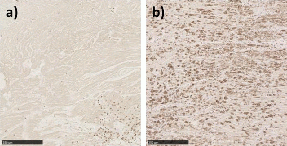
Results: There was no difference in inflammatory markers from heart tissue in the 105d gestation fetuses after myocardial infarction compared to control sham animals. However, there was a significant increase in the expression of inflammatory markers in the infarct tissue of 6 month old lambs compared to controls. When comparing the infarct area to the border and remote zone, there was a significant downregulation of pro-inflammatory cytokines such as IL-1β, IL-6 & TNF-α in the fetal infarct area with the opposite expression pattern occurring in the lambs. In addition, miRNAs involved in mediating the inflammatory response such as miR-125b and miR-23b were significantly downregulated in the lamb infarct compared to remote zone. Immunohistochemistry staining revealed differing localization of macrophages in the infarcted tissue area between the age groups (Figure).
Immunohistochemistry staining for the macrophage marker IBA-1 in the fetal (a) and the six month old (b) sheep heart infarcted areas.
Conclusion: These data indicate that the immune response to cardiac injury in the fetus is different to that of the adult, and the reduced inflammatory response seen in fetal hearts may contribute to the documented regenerative capacity in utero.