APPS November 2002 Meeting Abstract 1126


MEASUREMENT OF AREA CHANGES IN HUMAN AIRWAY SMOOTH MUSCLE CELLS USING QUANTITATIVE PHASE MICROSCOPY

Claire L. Curl1, Trudi Harris2, Akram A. Kabbara1, Brendan E. Allman4, Ann Roberts3, Keith A. Nugent3, Peter J. Harris1, Alastair G. Stewart2, Lea M.D. Delbridge1, 1 Department of Physiology, 2 Pharmacology, 3 School of Physics, University of Melbourne, Vic, 4 Imaging Division, IATIA Limited, Melbourne, Vic 3128.


0x08 graphic

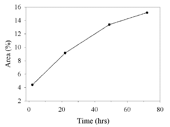
Fig 1: Area measurements of human airway smooth muscle cells calculated using quantitative phase microscopy


Enhanced visualisation of translucent cellular specimens may be achieved by detection of the phase shift in light traversing the sample. Quantitative Phase Microscopy (QPM) is a recently developed computational approach that provides quantitative phase measurements from images captured using a bright-field microscope without phase or interference contrast optics. An in-focus bright field image is acquired together with one positive and one negative defocused image, and an algorithm is then applied to produce a phase map(1). QPM was used to estimate the area of human airway smooth muscle cells, tracking the changes in growth over the period of a week. Cells were obtained by collagenase and elastase digestion of smooth muscle in airways from lung transplant resection patients. The resulting single cell suspension was washed in phosphate buffered saline and incubated in plastic culture dishes at 37oC in Dulbeccos Modified Eagles Media. Cells were imaged using an inverted Zeiss Axiovert 100 microscope fitted with a Zeiss LD-Achroplan (x10, 0.3 NA) objective and a Coolsnap fx CCD camera (Photometrics, USA). Phase measurements were made using QPm® software (v1.90 IATIA Ltd, Australia). When compared with intensities observed in bright-field images, the calculated phase map data demonstrated markedly enhanced contrast between cells and background and allowed determination of a more robust threshold point based on maximum intensity gradients. The total cell area was estimated using Image-Pro Plus software (v3.0 Media Cybernetics, USA). Figure 1 shows changes over 72 hours in the area of the culture dish occupied by cells. Using QPM, translucent cells can be visualised with improved cell boundary definition allowing precise and reproducible measurements of cell area, demonstrating that quantitative phase imaging can provide a rapid and non-destructive approach for measurement of cellular morphology.

(1) Delbridge LMD, Kabbara AA, Bellair C, Allman BE, Nassis L, Roberts A, Nugent KA. Today's Life Science. 2002;14:28-32


Programme Next